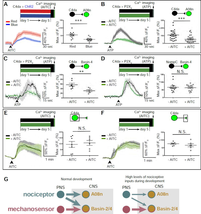
Figure 3. The sensory-input-induced plasticity of larval nociceptive circuit is pathway-specific.
(A) Optogenetic activation of C4da neurons during development inhibits C4da-to-A08n transmission. C4da neurons in mature larvae were stimulated by AITC for Ca2+ imaging. Max ΔF/F0 for individual A08n neurons are plotted on the graph and used for statistical analyses (i.e., each dot indicates one A08n). n = 12 neurons (from 6 larvae) per group.
(B) Stimulation of C4da neurons by AITC (5 mM) during development inhibits C4da-to-A08n transmission. C4da neurons in mature larvae were stimulated by P2X2 for Ca2+ imaging. n =26 (from 13 larvae) and 28 neurons (from 14 larvae) in “-AITC” and “+AITC”, respectively.
(C) Stimulation of C4da neurons by AITC (2.5 mM) during development inhibits C4da-to-Basin4 transmission. Because the responses of Basin-4 neurons are highly variable within each VNC (data not shown) (Jovanic et al., 2016), the average of Max ΔF/F0 of 10 Basin-4 neurons from abdominal segments 3 to 7 in each VNC were calculated to represent Basin-4 activity in each VNC (i.e., each dot indicates one larva). n = 7 and 6 larvae in “- AITC” and “+ AITC”, respectively.
(D) Treating larvae with AITC (2.5 mM) during development does not alter the mechanosensor-to-Basin4 transmission. n = 14 larvae per group.
(E–F) Larvae raised in environments with and without AITC (2.5 mM) have similar levels of noxious stimulation-induced calcium responses in C4da somas (E) and axon terminals (F). (E) n = 6 and 7 neurons (4 larvae per group) in “- AITC” and “+ AITC”, respectively. (F) n = 6 neurons from 3 larvae per group.
(G) Schematics showing the pathway-specificity of nociceptor-input-induced plasticity in synaptic connections between larval nociceptive and SONs in the circuit. Left: Circuit diagram of connections under normal developmental conditions. Right: High levels of nociceptive input specifically suppresses synaptic transmission from nociceptors to SONs.
(See also Figure S4.)

