
Figure 1.
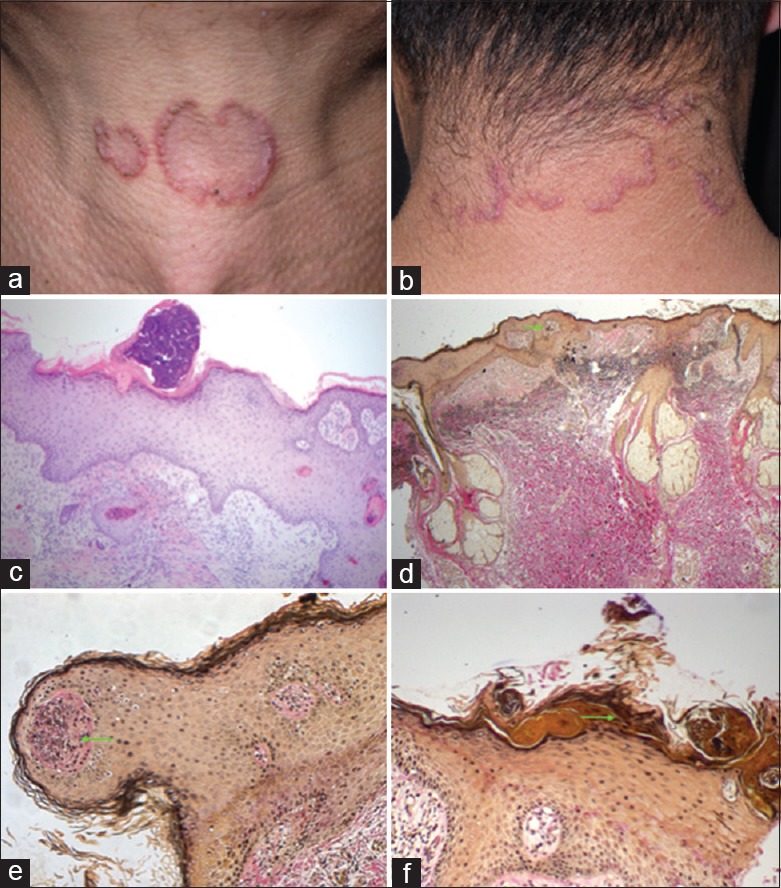
Representative images of the patient. D-penicillamine-induced elastosis perforans serpiginosa. (a and b) Physical examination showed multiple reddish-brown keratinized papules coalescing to form serpiginous plaques in the anterior and posterior of the neck. Scale and horny material can be observed in the center of the papules. (c) Epidermal hyperplasia with parakeratosis and dyskeratosis. Fiber necrosis can be observed in superficial dermis and especially in the stratum corneum. Lymphocyte, neutrophil infiltration is visible in focal location (Hematoxylin and Eeosin, ×100). (d) Elastic fibers mainly of the reticular dermis are coarser. Its perpendicular budding from the epidermis surface can be observed (arrow, Verhoeff-van Gieson staining, ×40). (e and f) Broken elastic fibers extruded through epidermis (arrow, Verhoeff-van Gieson staining, ×200).