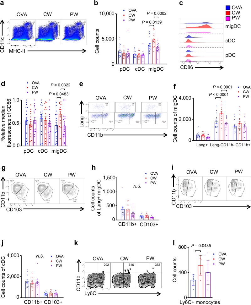
Figure 2. The effect of NIR laser adjuvant on DCs within the skin draining lymph nodes.
DCs in skin-draining lymph nodes (skin-dLN) were processed and stained for multi-parameter flow cytometry 24 hours after intradermal vaccination with 40 µg Alexa Fluor-488-labeled OVA with or without one minute CW or PW 1064 nm NIR laser treatment. (a) Representative gates of plasmacytoid DCs (pDC), classical lymphoid tissue-resident DCs (cDCs), and migratory DCs (migDC); numbers indicate percent of total lymphocytes. (b) Cell counts. (c) Representative histograms of CD86 expression. (d) Median fluorescent intensity of CD86 expression for pDC, cDC, and migDC population. (e) Representative gates of migDC subsets. (f) Cell counts of migDC subpopulation within skin-dLN. (g) Representative gates of Lang+migDC subsets, numbers indicate percent parent. (h) Cell counts of Lang+migDC subpopulation within skin-dLN. (i) Representative gates of cDC subsets, number representing percent parent. (j) Cell counts of cDC subpopulation within skin-dLN. (k) Representative gates of CD11b+Ly6C+ monocytes. (l) Cell counts of CD11b+Ly6C+ monocytes within skin-dLN. Data were analyzed with (b, f, h, j and l) two-way ANOVA followed by the Tukey's honestly significant difference (HSD) tests or (d) Kruskal-Wallis with Dunn’s correction for multiple comparisons. Experimental and control groups: (a–j) n = 16, 16, 17, (k–l) n = 6, 6, 7, for OVA i.d., OVA i.d. + CW 1064 nm, OVA i.d. + PW 1064 nm, respectively. Data are derived from three independent experiments.

