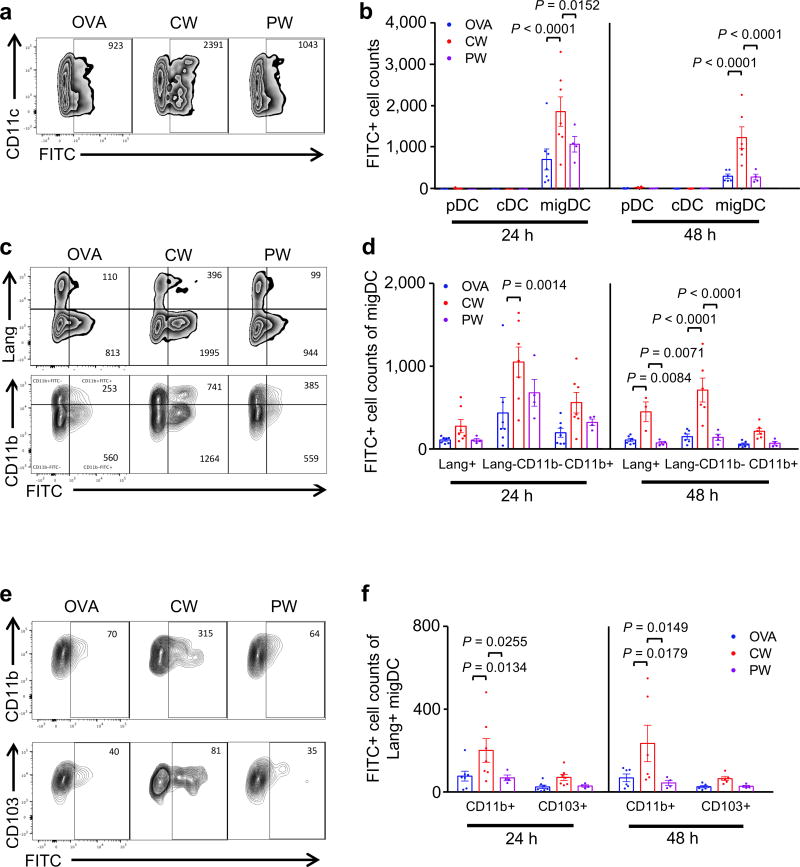
Figure 4. The effect of NIR laser on emigration of migDC subsets.
Mice were shaved, depilated, and painted with 1% FITC solution on the flank skin 4 hours before vaccination with 40 µg of OVA with or without NIR laser treatment. At the times indicated, single-cell suspensions from skin-dLN were labeled and analyzed based on surface markers and FITC fluorescence by flow cytometry. (a) Representative gates of FITC+ migDC emigrating into the skin-dLN after FITC painting. (b) Cell counts of in the skin-dLN after FITC painting. (c) Representative gates of migDC subsets. (d) Cell counts of migDC subpopulation within skin-dLN after FITC painting. (e) Representative gates of Lang+migDC subsets. (f) Cell counts of Lang+migDC subpopulation within skin-dLN after FITC painting. (b, e, f) Two-way ANOVA followed by the Tukey's HSD tests. Experimental and control groups: n = 6–7, 6–7, 4 for vaccine i.d., vaccine i.d. + CW 1064 nm, vaccine i.d. + PW 1064 nm, respectively. Data are derived from three independent experiments.

