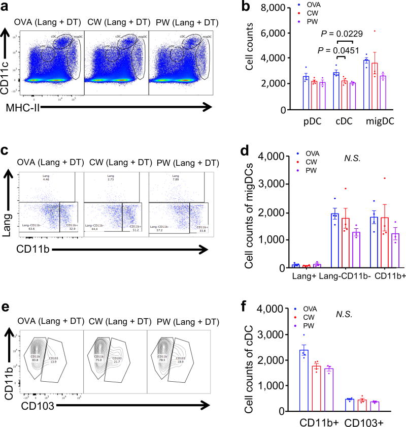
Figure 5. Depletion of Lang+ DCs removes CW laser induced population changes.
Lang−DTR/GFP mice were treated with diphtheria toxin 1 day prior to four intradermal injections of 10 µg of A488-labeled ovalbumin (OVA) with or without laser adjuvant illumination. DCs in skin-dLN were processed and stained for multi-parameter flow cytometry 24 hours after intradermal vaccination with 40 µg Alexa Fluor-488-labeled OVA with or without one minute CW or PW 1064 nm NIR laser treatment. (a) Representative gates of plasmacytoid DCs (pDC), classical lymphoid tissue-resident DCs (cDCs), and migratory DCs (migDC); numbers indicate percent of total lymphocytes. (b) Cell counts. (c) Representative gates of migDC subsets, numbers indicate percent parent. (f) Cell counts of migDC subpopulation within skin-dLN. (i) Representative gates of cDC subsets, number indicating percent parent. (j) Cell counts of cDC subpopulation within skin-dLN. (f, j) Data were analyzed with two-way ANOVA followed by the Tukey's honestly significant difference (HSD) tests. Experimental and control groups: (a–j) n = 4, 4, 3 for OVA i.d., OVA i.d. + CW 1064 nm, OVA i.d. + PW 1064 nm, respectively.

