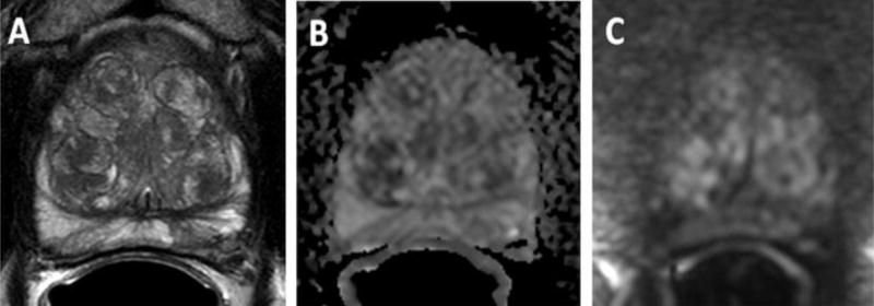
Figure 2.
66-year-old male with a serum PSA=10.8ng/ml. Pre-MRI biopsy revealed Gleason 3+3 prostate cancer (2% core involvement) in left apical PZ. mpMRI revealed no focal lesions but extensive BPH changes. Axial T2W MRI (A), ADC map (B) and b2000 DW MRI (C) shows no focal lesion. MRI excluded presence of a clinically significant lesion in this patient and patient proceeded to active surveillance. (Figure courtesy of Baris Turkbey, MD, National Cancer Institute)

