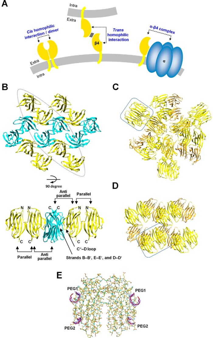
Figure 1.
Arrangements of the mouse/human β4 subunit extracellular domain molecules in the monoclinic, cubic, and hexagonal crystal forms. A, schematics of the cis and trans homophilic interactions of β4 and the α–β4 complex. The plasma membranes are colored gray. The cis homophilic interaction of β4 and the α–β4 complex occur on the same cell. Additionally, β4 can form a trans homophilic interaction between two different cells. B, arrangement of the mouse β4 molecule in the monoclinic crystal form (PDB code 5AYQ). The β4 molecules are colored yellow and cyan. Molecules with the same color are parallel to each other, and the yellow molecules are oriented anti-parallel to the cyan ones. Shown are a top view (top panel) and side view of the enclosed region (bottom panel). C and D, arrangements of the mouse β4 molecules in the cubic crystal form (C) and the human β4 molecules in the hexagonal crystal form (D). One of the pairs of β4 molecules interacting with each other in the parallel arrangement is enclosed. E, electron densities of the polyethylene glycol fragments. Shown is a 2Fo-Fc composite omit map (contoured at 1.5 σ) of the polyethylene glycol fragments (PEG1 and PEG2) in the hexagonal form.

