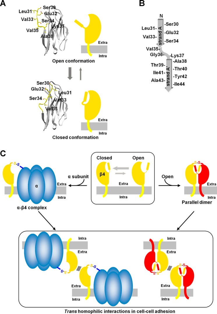
Figure 7.
Schematics of β4. A, Open (top panel) and closed (bottom panel) conformations. The plasma membranes are colored gray. The β4 protein may accommodate the hydrophobic residues in its own hydrophobic pocket to form a monomer in a closed conformation. B, topology diagram of strand A/A′. C, the trans homophilic interactions of the parallel dimer and the monomer of β4 in cell–cell adhesion. The closed and open conformations of β4 are likely to exist in equilibrium, and the parallel dimer could be readily formed when two monomers in the open conformation meet each other. The monomeric β4 can associate with the α subunit. Both the α-β4 complex and parallel dimer could exhibit cell–cell adhesion.

