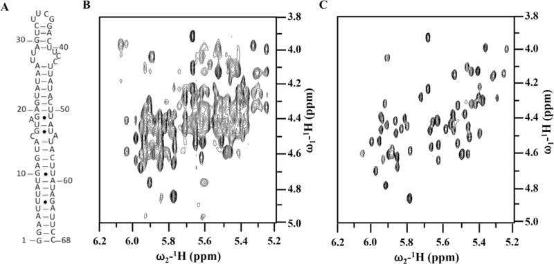
Fig. 4. Effect of site-specific ribose deuteration on the quality of the NMR spectra of an RNA thermometer.

(A) secondary structure of the ‘core’ thermometer. (B) and (C) are 2D 1H–1H NOESY spectra recorded in D2O for completely protonated (B) or with H6/H8, H1′, H2′, D3′, D4′, D5′/D5′ and D5 ribose deuteration, respectively, at 25 °C and 600 MHz. Spectral simplification is remarkable.
