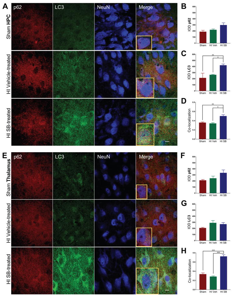
Figure 7. SB505124 stimulates association of autophagic markers LC3 and p62 following HI injury at P6 in the hippocampus and thalamus.
One week after HI injury at P6, samples of the injured forebrain were analyzed by immunofluorescence. SB505124 or vehicle was administered via osmotic pump beginning at 3 days after injury and maintained for 4 days prior to intracardiac perfusion. 30μm sections were stained with anti-p62 (red), anti-LC3 (green), and anti-NeuN (blue) markers. (7A, E) Hippocampal and thalamic neurons in the ischemic penumbra of injury, respectively. Insets depict representative cells enlarged an additional 2X. (7B, C) Integrated densities of p62 and LC3 fluorescence of Sham, Vehicle-treated HI, and SB-treated HI groups in each respective region. (7D, G) Manders’ colocalization coefficient (M1) for the fractional overlap of p62 signal in compartments containing LC3 signal for the cortex and white matter (n=3–5). Data are presented as means ± SEM *p<0.05, **p<0.001, ***p<0.0001 by ANOVA followed by Tukey’s post hoc test). Scale bars in merged image represent 10 μm.

