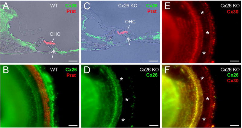
Fig. 1.
Deletion of Cx26 in the cochlea in Cx26-Prox1 cKO mice. A–B: Immunofluorescent staining for Cx26 (green) and prestin (red) in WT mice with the cochlear cross-section and whole-mounting preparation. Outer hair cells (OHCs) are visualized by prestin labeling. A white arrow indicates Deiters cell (DC) and outer pillar cell (OPC) area. C–F: Immunofluorescent staining of the cochlea for Cx26 and prestin in the Cx26-Prox1 cKO mice. A white arrow in panel C indicates no Cx26 labeling in the DC and OPC area. White asterisks in panel D–F represent OHC-DC-OPC area in the whole-mounting preparation, in which Cx26 labeling is absent while Cx30 labeling remains. Scale bar = 50 µm.

