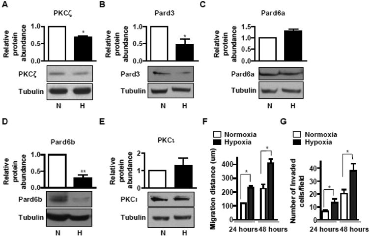
Fig 1. Hypoxia-mediated downregulaion of PKCζ/Pard3/Pard6 correlates with the induction of migration and invasion in non-small-cell lung carcinoma cells.

A-D) A549 cells were exposed to normoxia (N) or hypoxia (H, 1.5% O2) for two days. The whole cell lysates were prepared and aliquots with the same amount of protein were subjected to SDS-PAGE followed by Western blot analysis for PKCζ (A), Pard3 (B), Pard6a (C), Pard6b (D), and PKCι (E). The amount of tubulin was used as control for equal loading. Data were expressed as mean ± SEM. n = 4. *, p < 0.05; **, p < 0.01. F) A549 cells were cultured on 35-mm dishes overnight to reach confluence. Wounds were created by scratching a straight line with a 250-μl tip. After 24 hours or 48 hours of incubation in normoxia or hypoxia (1.5% O2), the widths of the wounds were measured and the difference between the width before and after migration was presented as the migration distance. Data were expressed as mean ± SEM. n = 5. *, p < 0.05. G) A549 cells were cultured on BD Matrigel invasion chambers for 24 or 48 hours in normoxia or hypoxia. Invaded cells were stained and the number of invaded cells in each field was counted under microscopic fields at 200x magnification. Experiments were carried out in triplicates and repeated three times. The results were compared to that of A549 cells and data are expressed as mean ± SEM. *, p < 0.05.
