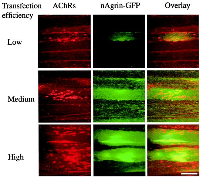
Figure 2.
Recombinant neural agrin and accompanying AChR aggregates in transfected SOL muscles. SOL muscles were denervated and injected with solutions of GFP-tagged neural agrin cDNA of low, medium, or high transfection efficiencies, as indicated. Eight days later, the muscles were removed, teased into bundles, incubated with Rh-BuTx, and examined with confocal microscope. Note the increase in the amount of recombinant GFP-tagged neural agrin (green fluorescence) and the number of AChR aggregates (red) on transfected (see overlay) and adjacent muscle fibers for each increase in transfection efficiency. (Scale bar, 50 μm.)

