
Figure 3. F nucleatum regulates miR21 expression in CRC.
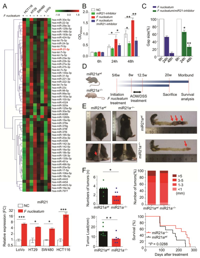
A) Microarrays were performed to identify the differentially expressed miRNA between F nucleatum-treated and control cells. The miR21 expression was consistently up-regulated in different CRC cells after F nucleatum treatment (*P < 0.05, ***P < 0.001, unpaired Student’s t test). B and C) CCK-8 cell viability assay and wound healing assays illustrated that inhibition of miR21 completely abolished F nucleatum’s effect on cell growth and invasion(*P < 0.05, **P < 0.01 by unpaired Student’s t test). D) Both miR21 WT and knockout (KO) mice were initially administrated F nucleatum and then subjected to AOM/DSS treatment. At 20 weeks, mice were sacrificed for various experiments. E) The left representative image showed miR21 KO decreased the bloody diarrhea rates during the F nucleatum infection (n = 7 per group), and the right image showed a representative colon of the miR21 KO or wild type mice. F) The colorectum in miR21 KO mice had fewer tumor numbers, smaller size and less tumor load compared to WT mice (n = 7 per group,*P<0.05, Mann-Whitney U test). Furthermore, miR21 KO mice had longer survival rates than WT mice (n = 10 per group by log-rank (Mantel-Cox) test). These results are representative of at least three independent experiments. Results represent means values ± SD. The red arrows indicated the positively stained cells.