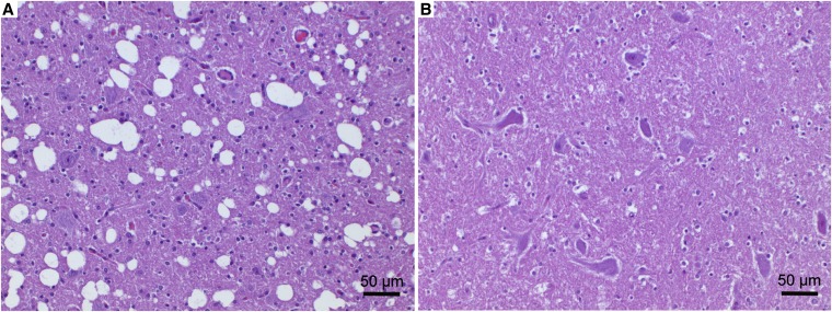
Figure 2.
Histopathology of a cerebellar nucleus. (A) Malinois dog MA162 with spongy degeneration and (B) nonaffected control dog. The affected Malinois puppy (A) showed a prominent vacuolation of the neuropil with large numbers of clearly defined and empty vacuoles of varying size and gliosis. Hematoxylin and eosin stain.

