Figure 1.

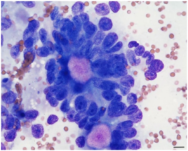
Fine-needle aspirate cytology from the right ovary. Epithelial cells arranged in acinar-like structures with bright eosinophilic granular material in the middle (Call–Exner bodies); × 40 oil objective, Modified Wright’s stain. Bar = 10 µm

Fine-needle aspirate cytology from the right ovary. Epithelial cells arranged in acinar-like structures with bright eosinophilic granular material in the middle (Call–Exner bodies); × 40 oil objective, Modified Wright’s stain. Bar = 10 µm