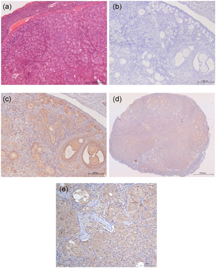
Figure 2.
(a) Haematoxylin and eosin-stained section of tissue of the ovarian neoplasm showing a region where the neoplasm is encapsulated. (b–e) Anti-Müllerian hormone immunoreactivity. (b) Antibody-negative control section of tissue from a healthy ovary. (c) Anti-Müllerian hormone immunoreactivity identified in primordial, primary and secondary ovarian follicles from a healthy ovary. (d) × 50 magnification, (e) × 200 magnification image demonstrating anti-Müllerian hormone immunoreactivity identified within the neoplastic population of granulosa cells

