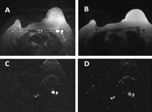
Figure 1.

A 62 Year-Old Woman with Breast Cancer. A, STIR shows two enlarged left axillary LNs displaying round shape and attenuated fatty hila; B, T1W non-Fat Sat; C, DWI shows high signal intensity; D, ADC displays metastatic LNs with restricted diffusion and ADC value = 0.872 ×10⁻³ mm2/s
