
Figure 9.
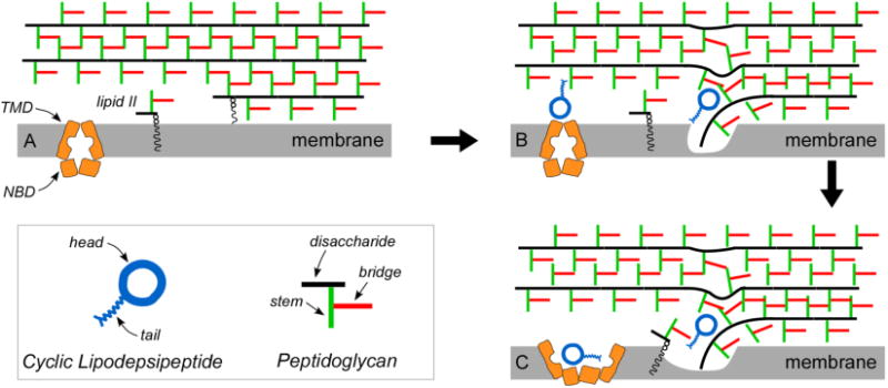
(A) Schematic drawing of the peptidoglycan layers near the bilayer of whole cells of S. aureus. (B) The headgroup of plusbascin A3 inserts near a bridge (red) in the hydrophobic region of the peptidoglycan, and the tail displaces the glycan chain (black) which disorganizes the bilayer. The head-and-tail combination prevents extension of the nascent peptidoglycan by addition of lipid II. (C) A second plusbascin A3 in the hydrophilic region of the cell wall (see previous panel) is shown blocking the function of an ATP-binding cassette transporter, possibly resulting in proximity of purines in ATP to both plusbacin A3 head and tail. Abbreviations: TMD, transmembrane domain; NBD, nucleotide-binding domain.