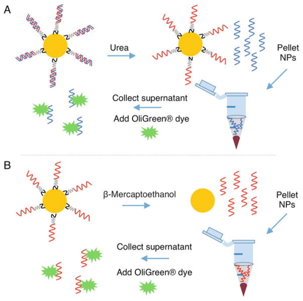
Fig. 2.
Schematic representing the procedure to quantify siRNA duplexes on nanoparticles (NPs). (a) Antisense RNA strands (blue) are dehybridized from sense RNA strands (red) on nanoparticles by incubating in 8 M urea at 45 °C. The sense-loaded nanoparticles are pelleted by centrifugation, and the supernatant containing the antisense strands is collected. Antisense RNA strands within the supernatant are measured using components of the Quant-iT™OliGreen® kit. (b) Sense RNA strands (red) are removed from the nanoparticles’ surfaces by breaking the gold–thiol bond with β-Mercaptoethanol. The nanoparticles are pelleted by centrifugation, and the supernatant containing the sense RNA strands is collected for analysis of RNA content with the Quant-iT™ OliGreen® kit

