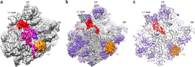
Figure 1.
The structure of the pre-60S Nmd3-TAP ribosomal particle. (a,b) Low-pass-filtered (a) and B-factor-sharpened (b) cryo-EM density maps of the pre-60S particle, shown as surface representations. Lsg1, Nmd3, the N-terminal domain of Nmd3 (Nmd3-N; violet in a) and Tif6 are indicated by color-coding. Ribosomal proteins are shown in purple in b and c. (c) A representation of the final atomic model. The dashed circle indicates the PTC. SB, P0 stalk base.

