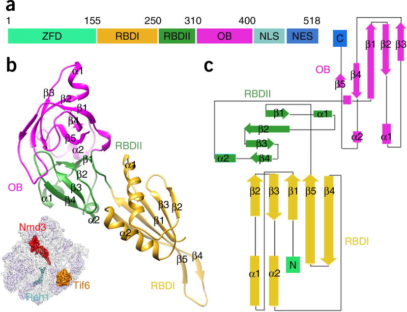
Figure 2.
The structure of Nmd3. (a) Schematic illustration of Nmd3 domain organization. ZFD, zinc-finger-containing domain; NLS, nuclear localization sequence; NES, nuclear export signal. (b) The atomic model of Nmd3, with individual domains (RBDI, RBDII and OB) indicated by color-coding. The orientation of Nmd3 on the pre-60S particle is shown in the illustration on the lower left. (c) Domain topology of Nmd3.

