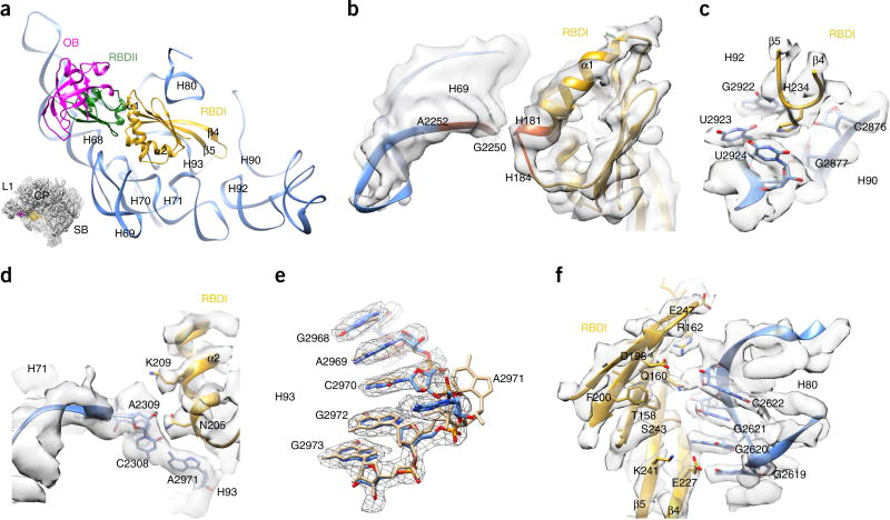
Figure 3.
Interaction of Nmd3 RBDI with 25S rRNA. (a) Binding position of Nmd3 on the pre-60S particle. Different Nmd3 domains are indicated by color-coding. The orientation is represented in the illustration on the lower left. (b) Zoomed-in view of the interaction between the α1 helix of RBDI and H69. (c) H234 is situated in the PTC and interacts with G2922, U2923, U2924 and C2876 of 25S rRNA. (d) Zoomed-in view of the interaction between the α2 helix of RBDI and H71–H93. It is likely that A2971 of H93 forms a noncanonical base pair with C2308 of H71. (e) Conformational difference between A2971 in the pre-60S map and in its mature state. Coordinates of the mature state (tan) are from the crystal structure of the yeast 80S ribosome (PDB 3U5E)27. (f) Extensive interactions between the β-sheet surface of RBDI and H80.

