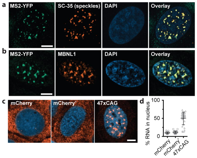
Figure 3. 47xCAG RNA is retained within the nucleus and co-localizes with nuclear speckles.

(a) Immunofluorescence images depicting co-localization of 47xCAG RNA foci (MS2-YFP) with nuclear speckles (SC-35). Nuclei are counterstained with DAPI. (b) Similar to (a), staining for MBNL1. (c, d) FISH images (c) and relative RNA abundance per nucleus (d), in cells expressing MS2-tagged 47xCAG, a control coding sequence (mCherry) or its reverse complement (mCherry′). Scale bars, 5 μm. Error bars, mean ± s.d. Data are representative of ≥ 3 independent experiments.
