Figure 3.

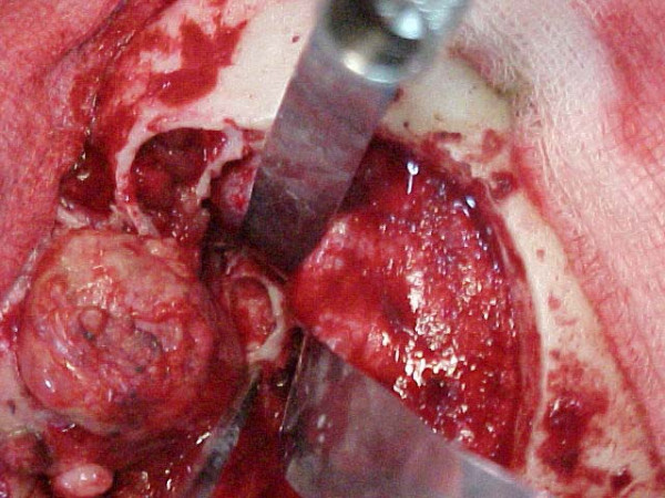
Intracranial portion of the tumor. Dura being retracted after the craniotomy and visualization of the intracranial portion of the tumor. The tumor core has been drilled. The shell was subsequently nibbled away.

Intracranial portion of the tumor. Dura being retracted after the craniotomy and visualization of the intracranial portion of the tumor. The tumor core has been drilled. The shell was subsequently nibbled away.