Figure 6.

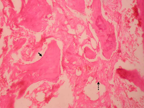
Microphotograph of the tumor. Microphotograph (250X) of the specimen in H & E stain showing new osteoid lined by plump osteoblasts (solid arrow) in a highly vascularised stroma (broken arrow).

Microphotograph of the tumor. Microphotograph (250X) of the specimen in H & E stain showing new osteoid lined by plump osteoblasts (solid arrow) in a highly vascularised stroma (broken arrow).