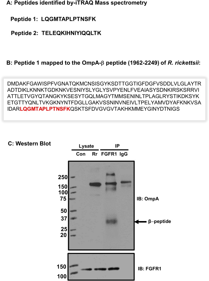
Fig 4. FGFR1 interacts with β-peptide of OmpA.
Confluent ECs were infected with R. rickettsii and FGFR1 was immunoprecipitated from the total protein lysates. The samples were subjected to mass spectroscopic analysis using isobaric tag for relative and absolute quantitation [iTRAQ] method [described in materials and methods]. (A): The peptides interacting with FGFR1 were identified as peptide 1 and 2. (B): The location of the peptide 1 within the beta peptide sequence of OmpA is shown in red. (C): FGFR1 was immunoprecipitated (IP) from R. rickettsii-infected ECs and samples were subjected to SDS-PAGE and Western blotting using rickettsial OmpA antibody. Mouse IgG was used as the control. The blot was also probed with an FGFR1 antibody to demonstrate that the immunoprecipiatation was successful. A representative blot from three independent experiments is shown.

