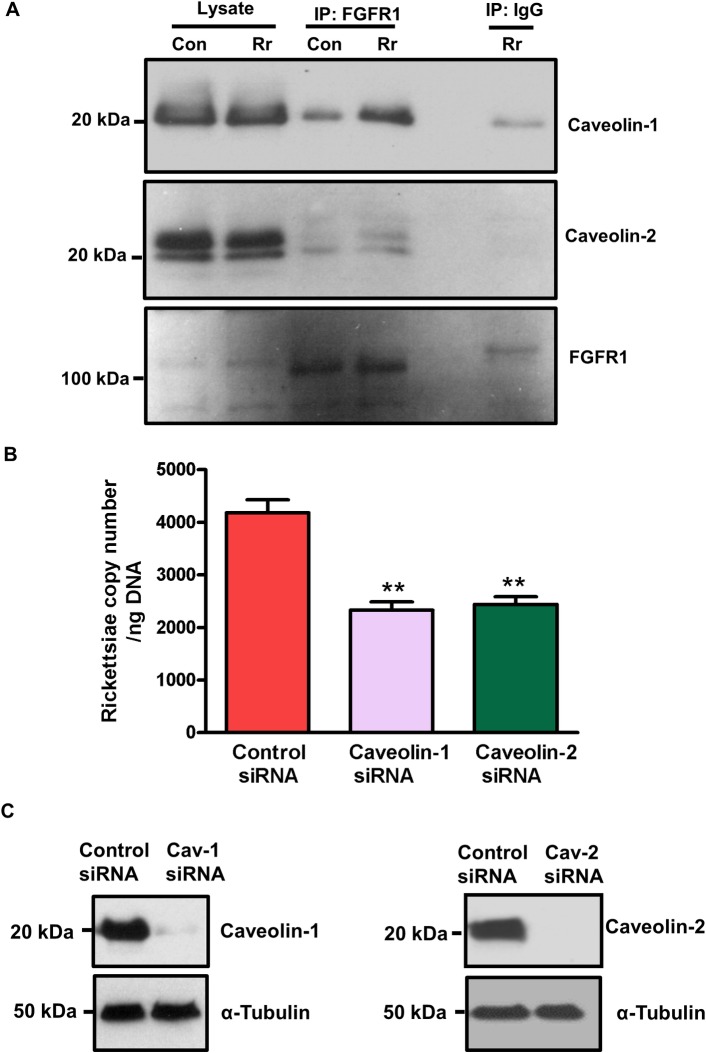
Fig 5. FGFR1 interactions with caveolin-1 and caveolin-2.
(A): Confluent ECs were infected with R. rickettsii. At 1 hour post-infection, the cell lysates were prepared, FGFR1 was then immuno-precipitated using an FGFR1-specific antibody. Mouse IgG was used as a negative control. Samples were subjected to SDS-PAGE and Western blotting using antibodies against caveolin-1, caveolin-2 and FGFR1. (B): ECs were transfected with either control, caveolin-1 or caveolin-2 siRNA for 72 hours and then infected with R. rickettsii for 1 hour. Rickettsial copy number was measured by q-PCR using the OmpA primer pair. The asterisks represent a significant change (p≤ 0.001). The data are presented as the mean ± SE of three independent experiments. (C): caveolin-1 (cav-1) and caveolin-2 (cav-2) expression were measured by Western blotting to demonstrate the functionality of siRNAs used in our experiments.

