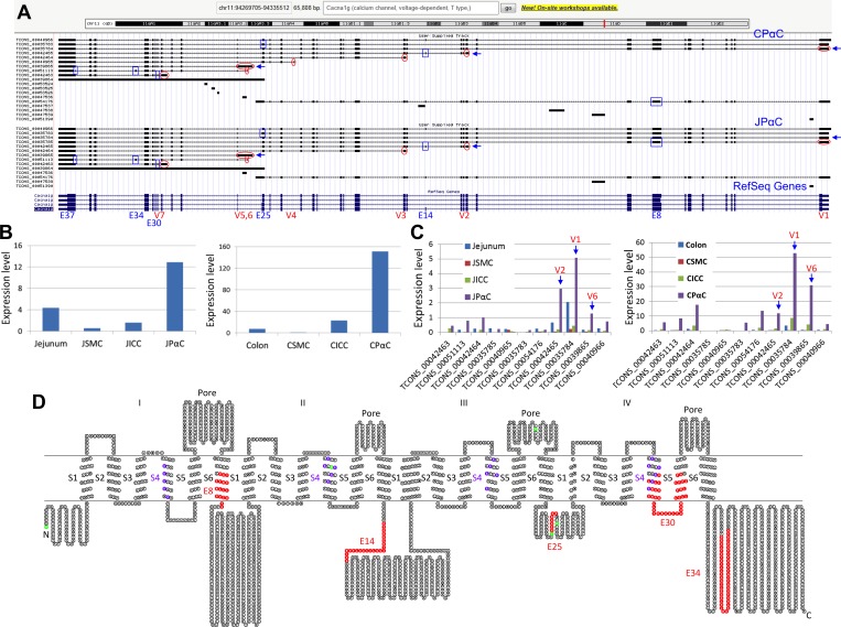
Fig 3. Identification of mutiple Cacna1g trancritional variants.
(A) A genomic map view of Cacna1g variants expressed in JPαC and CPαC. Seven alternative initial exons (V1-7) are circled in red and six differentially spliced exons (E8, E14, E25, E30, E34, and E37) are boxed in blue. (B) Expression (FPKM) levels of total Cacna1g mRNAs in JPαC and CPαC. (C) Expression levels of Cacna1g transcriptional vaiants in JPαC and CPαC. (D) A topological map of CACNA1G variants. Each circle denotes a single amino acid. Colors on amino acid sequence show distinct regions and domains. Red represents missing, or inserted, peptides from differentially spliced exons. Green represents start codons found in differentially spliced variants. Six transmembrane domains (S1-6) and a pore region are shown.

