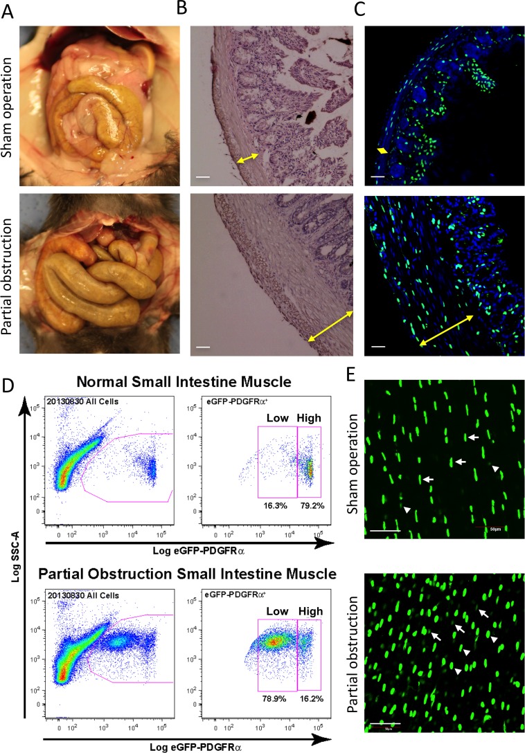
Fig 4. Increased PDGFRα+ cells in hypertrophic smooth muscle.
Hypertrophic tissue was surgically induced for ~2 weeks by placing a small silicone ring on the distal ileum of transgenic PDGFRα-eGFP mice to partially obstruct normal peristaltic movement. (A) Gross images of GI tract in sham and obstruction surgeries. (B) Representative H&E staining of jejunal cross sections from sham control and partially obstructed mice. Hypertrophied jejunum contained significantly thicker circular and longitudinal muscle layers compared to a sham control. Scale bar: 50 μm. (C) Representative confocal laser scanning images of jejunal cross sections from sham operation control and partial obstruction mice showing nuclear eGFP expression in PDGFRα+ cells and DAPI (blue) counterstained in the cells. Scale bar: 50 μm. (D) Two populations (eGFPhigh and eGFPlow) of primary PDGFRα+ cells from hypertrophic jejunum identified by flow cytometry. Note that eGFPhigh PDGFRα+ cells are significantly increased in partial obstruction smooth muscle. (E) A z-stack image, obtained through confocal microscopy, of whole-mount jejunum muscularis from the partial obstruction (bottom) and sham operation control (top) showing eGFPhigh (arrow heads) and eGFPlow (arrows) PDGFRα+ cells. Scale bar: 50 μm.

