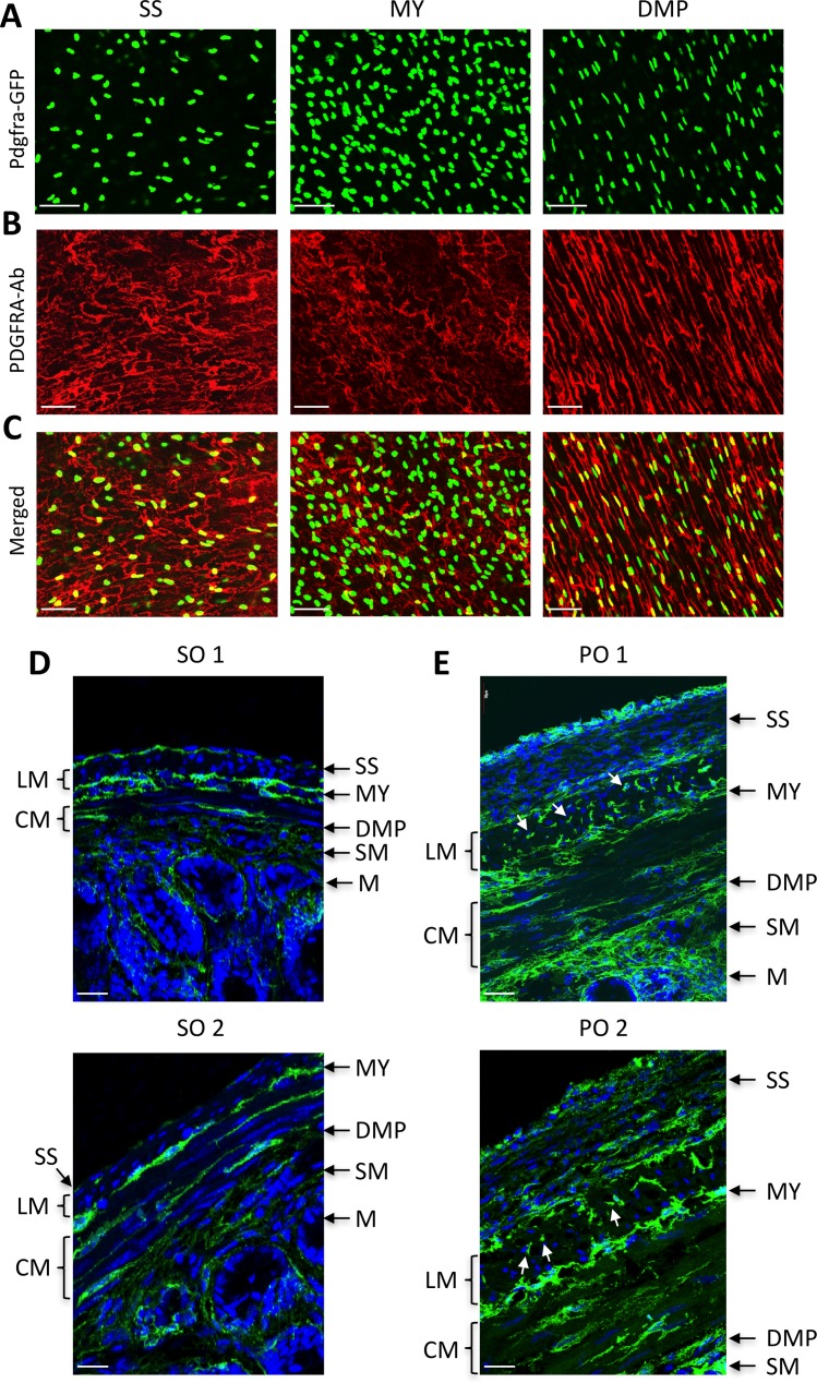
Fig 5. Identification of PDGFRα+ cell subpopulations dedifferentiated in hypertrophic smooth muscle.
(A-C) Confocal section images of PDGFRα+ (Pdgfra-eGFP+) cell subpopulations identified in the subserosal layer (SS), myenteric region (MY), and deep muscular plexus (DMP) with PDGFRA antibody (A), eGFP (B) and merged (C) in jejunum. (D and E) Cross section images of PDGFRα+ cell subpopulations (green) in sham operation (SO, D) and partial obstruction (PO, E). Proliferating PDGFRα+ cells are marked by arrows. LM, lonitudinal muscle; CM, circular muscle; SM, submucosa; M, mucosa. All scale bars are 50 μm.

