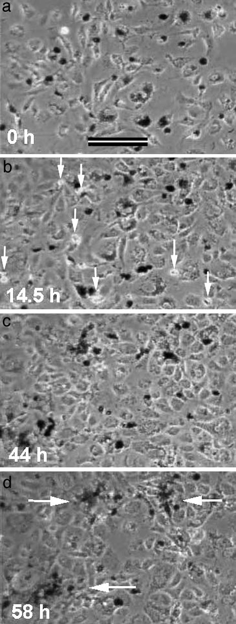
Fig. 3.
Time-lapse observation of the aggregation of hyperscattering cells mixed with particle-free 3T3x cells. The hyperscattering cells contained 1-μm latex particles and appear dark in phase contrast (low levels of 600-nm field illumination). White arrows in b point to several mitotic figures. White arrows in d point to final aggregates. The hyperscattering cells drew closer to each other long before they were able to make physical contact.

