Fig. 3.
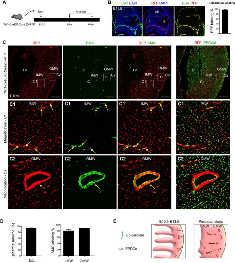
Early embryonic epicardial cells contribute to SMCs of the 2nd CVP. (A) A schematic figure depicting the experimental time-course shows tamoxifen induction (at E10.5) and endpoint analysis (at postnatal 8 weeks, P8w to P16w) of Wt1-CreER; Rosa26-RFP mice. (B) Following tamoxifen administration at E10.5, Wt1-CreER; Rosa26-RFP embryos were harvested at E11.5. Active WT1 expression was assessed by ESR immunostaining, and epicardial cell genetic labeling was determined by RFP. Nuclei were stained with DAPI. Labeling efficiency was calculated as the percentage of RFP + ESR + epicardial cells divided by the total number of ESR + epicardial cells. Scale bars, 100 µm. Li, liver; a, atrium; v, ventricle; oft, outflow tract. (C) Immunostaining of RFP, SMA, PECAM and DAPI on P10w Wt1-CreER; Rosa26-RFP heart sections. Smooth muscle cells (SMCs) of coronary arteries in both the inner (C1) and outer myocardium wall (C2) were derived from Wt1+ epicardial cells labeled at E10.5 (arrows). Scale bars, 0.5 mm. LV, left ventricle; IMW, inner myocardial wall; OMW, outer myocardial wall. Representative figure of 4 individual samples. (D) Quantification of panels in “C” shows the contribution of the Wt1-CreER lineage to the epicardium and SMCs. n = 4. (E) A hypothetical model illustrating epicardial cell (green) migration (black arrows) and establishment of EPDCs (red) in the compact myocardium at E10.5-E13.5 (left panel). Postnatally, a subset of these resident EPDCs (red) migrate (black arrows) from the OMW into the IMW to form the SMCs of the 2nd CVP.

