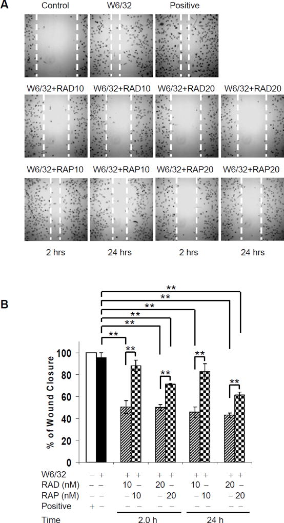
Fig. 6. Everolimus inhibits HLA I-mediated cell migration more potent than sirolimus.
A HAEC were grown in 35 mm culture dishes coated with 0.1% gelatin up to confluence. Quiescent cells were pretreated with 10 µg/ml of mitomycin C for 2hr to inhibit cell proliferation before being assayed for their ability to migrate. A scratch wound was created with a sterile 200-µl pipette tip. Wounded cells were pretreated with 10 or 20nM of everolimus or sirolimus at 10nM or 20nM for 2 or 24hr, and then were stimulated with 1.0 µg/ml of anti-HLA I mAb W6/32 for 24hr. EC were incubated in complete medium as positive control. Representative microscopy fields are shown. B, The distance between two edges was measured; migration rate was analyzed by calculating the distance of two front edges of class I-stimulated EC divided by the distance of two edges of control EC. C, HAEC were grown in 24-well plate coated with 0.1% gelatin up to 90% confluence. Quiescent endothelial cells were pretreated with or without everolimus or sirolimus for 2 or 24hr. Migration of HAEC was measured in a transwell insert system. HAEC pretreated with or without mTOR inhibitors were added to the upper chamber of insert and stimulated with mAb W6/32. HAEC treated with VEGF at 10ng/mL served as positive controls. After incubation for 16hr at 37°C, the cells on the upper surface of the membrane were removed with a cotton swab, and the migrated cells were fixed with methanol, stained with crystal violet, and three middle fields per insert were photographed with 10 × objective lens, and counted. Fluorescence 10x microscopy images are presented. D, The bar graph shows the mean ± SEM number of migrated cells. *p<0.05, **p<0.01, and ***p<0.001 were analyzed by one way ANOVA with Fisher’s LSD. Data represent at least three independent experiments. HAEC used in these experiments include CAR, CAS, and 5555.

