
Figure 5.
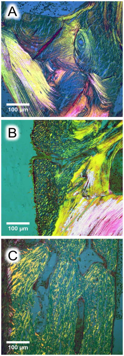
Histological images of cortical bone from transiliac biopsy samples (A and B: control, C: OI type V). The sections were stained by Goldner trichrome and observed in polarized light. (A) Ordered lamellar collagen organization in a healthy control (2 year-old child), (B) periosteal primary bone apposition in the same child viewed as disorganized collagen fibrils pattern abutting to the secondary remodeled bone with lamellar pattern, (C) collagen organization in a 6.8 year-old child with OI type V suggesting short range and randomly organized fibril throughout the cortex similarly to the periosteal primary bone apposition observed in B.