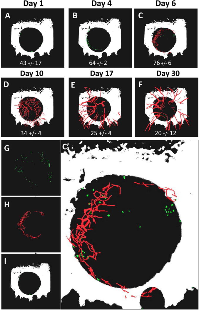
Figure 3. Mast cells accumulate at the edge of cranial defects within 24hrs of injury prior to marked angiogenesis at 1 week.

WT female C57BL/6 mice (n=3) underwent cranial defect window surgery, and MPLSM was performed after injection of Texas red dextran and FITC conjugated anti-MCPT5 antibody on days 1, 4, 6, 10, 17 and 30 post-op as described in Materials and Methods. 3D reconstruction of the multiphoton images acquired at 4× was performed with Amria, and images from a representative mouse with the number of mast cells (mean +/− SEM for the cohort) within the defect at each time point is shown (A–F) to illustrate the rapid accumulation of mast cells around the edge of the defect relative to angiogenesis over the course of bone defect healing. Individual renderings of the day 6 image (C) are shown to better illustrate the FITC-labeled mast cells (green in G), Texas red dextran labeled vasculature (red in H), and bone from second harmonic generation (white in I), and combined in a enlarged 3D rendering (C’).
