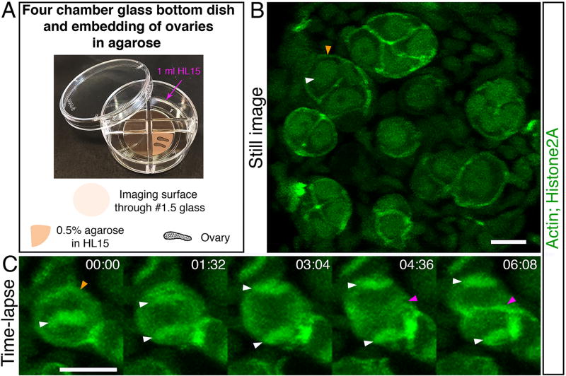
Figure 5. Live time-lapse imaging.
Ovaries of fish expressing Lifeact-GFP to label Actin and H2A-GFP to label chromatin via Histone2A [Tg (βact:Lifeact-GFP); Tg(h2afva:h2afva-GFP)] were collected and imaged live. (A). A four-chamber glass bottom dish used for mounting 27 cultured live ovaries for time-lapse imaging. (B) A still image from a time-lapse recording showing several 2 to 8-cell oogonial cysts. Image is a partial projection not showing all cells in each cyst. White arrowhead indicates chromatin detected by H2A-GFP labeling. Orange arrowhead indicates the cortex of the cell as detected by the Lifeact-GFP. Scale bar is 10 µm. (C) A dividing oogonial cell progressing from metaphase, to anaphase, and cytokinesis. Images are partial projections. Time is indicated in minutes (min). Scale bar is 10 µm. White arrowheads indicate the segregating chromosomes (H2A-GFP) from the metaphase plate and into individual daughter cells. The orange arrowhead indicates the cortex of the cell as detected by Lifeact-GFP at time 00:00 min. The magenta arrowhead indicates the appearance of a cleavage furrow in cytokinesis at time 04:36 min, and presumptive membrane between daughter cells at time 06:08 min Tg(βact:Lifeact-GFP) and Tg(h2afva:h2afva-GFP) were reported [33] [34], respectively.

