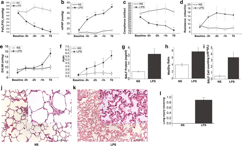
Fig. 1.

LPS impairs lung function and alveolar-capillary barriers. Pigs were allocated into intravenous saline and LPS group, respectively. Parameters of oxygenation, hemodynamics and lung mechanics were recorded at different time points from baseline to the end of the experiment (T0). BALF and lung tissues were collected at T0. PaO2/FiO2 (a) was measured by using blood samples from femoral artery on a blood gas analysis instrument. The value of mPAP (b) was gained by Swan-Ganz catheter implantation into jugular vein. Compliance (c) and resistance (d) were recorded by using Drager Evita 4 ventilator. EVLWI (e) and PVPI (f) were measured by PiCCO system at baseline and a series of time points until ALI was well established (T0). g Whole protein content in the BALF of pigs was detected by using BCA protein assay kit. h The ratio of wet/dry lung weight of saline control and LPS challenge pigs. i Whole-cell counts in the BALF collected from both groups of pigs. j and k were the representative histology sections of controls and LPS challenge pigs. HE sections are from one representative animal per group. l Lung injury scores were calculated according to ATS report which was in detail described in the “Methods.” Data points are expressed as mean ± SEM, N = 6 of each group. Statistical significance between saline and LPS group per point of measurement is shown as *P < 0.05; **P < 0.01; ***P < 0.001, respectively
