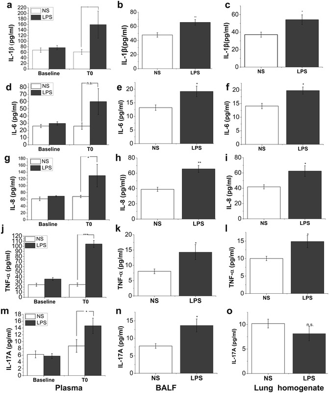
Fig. 2.

Concentrations of circulating and pulmonary inflammatory parameters. In LPS group, plasma samples were collected before LPS challenge (baseline) and at the time when ARDS model had been well established (T0). BALF and lung homogenates were also prepared at necropsy. Samples from saline control group were also obtained at the corresponding time points. The level of circulating and pulmonary IL-1β (a–c), IL-6 (d–f), IL-8 (g–i), TNF-α (j–l) and IL-17A (m–o) was detected by using commercial ELISA kits. Data are presented as mean ± SEM (N = 6 of each group). Compared with saline (NS) group, *P < 0.05; **P < 0.01; ***P < 0.001
