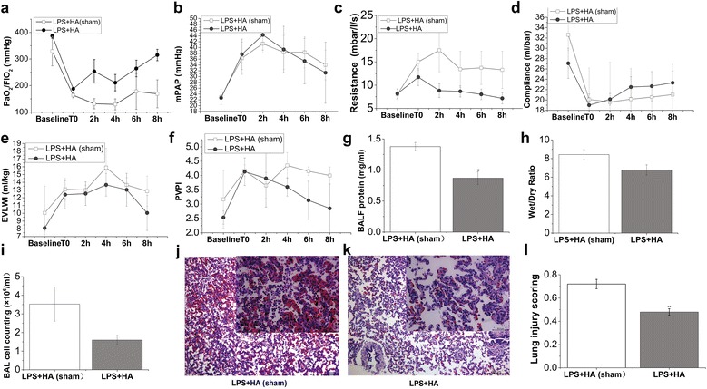
Fig. 3.

Effects of HA on oxygenation, lung mechanics and alveolar-capillary barriers. Pigs were challenged with LPS (50 μg/kg) intravenously to recapitulate the features of ARDS. When ARDS was well established (T0), animals received hemoadsorption (HA) or sham-hemoadsorption treatment for 3 h. Following another 5-h observation, animals were killed for BALF and lung tissue processing. At baseline, T0, and a series of time points during (2 h) and after HA (or HA-sham) treatment (4, 6 and 8 h), parameters of oxygenation and lung mechanics were recorded. PaO2/FiO2 (a) was measured by using blood samples from femoral artery on a blood gas analysis instrument. The value of mPAP (b) was gained by Swan-Ganz catheter implantation into jugular vein. Resistance (c) and compliance (d) were recorded by using Drager Evita 4 ventilator. EVLWI (e) and PVPI (f) were measured by PiCCO system at indicated time points (baseline, T0, 2, 4, 6 and 8 h following HA or HA-sham treatment). BALF protein (g) was assessed at 8 h after HA (or HA-sham) as described in “Methods”. h The ratio of wet/dry lung weight of animals in both groups was calculated by gravimetric method. i Whole-cell counts in the BALF collected from both groups of pigs were numerated by using a counting plate. j and k were the representative lung histology sections of pigs in LPS + HA (sham) and LPS + HA groups. HE sections are from one representative animal per group. l Lung injury scores were calculated according to ATS report which was in detail described in the “Methods.” Data points represent as mean ± SEM, N = 3–4 of each group and time point. Statistical significance between HA and HA-sham group per point of measurement is shown as *P < 0.05
