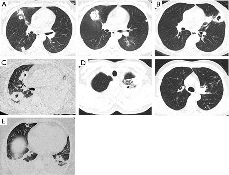
Figure 1.
Chest CT image using the lung window setting. (A) Chest CT image shows multiple changes in lesions and PCs with thick walls (“air crescent sign”); (B) chest CT demonstrates a PC with a thick wall; (C) chest CT image manifests multiple PCs with atelectasis; (D) chest CT image shows multiple changes in lesions with thick-walled PCs (“tree-in-bud sign”); (E) CT shows multiple thick-walled PCs with pleural effusions. PC, pulmonary cavity.

