
Figure 5.
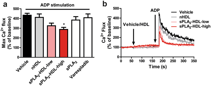
sPLA2-HDL inhibits Ca2+ flux in platelets. Baseline Ca2+ levels were measured by flow cytometry for 1 min and then platelets were treated either with vehicle, nHDL (50 µg/mL), sPLA2-HDL-low (50 µg/mL), sPLA2-HDL-high (50 µg/mL), sPLA2 or varespladib for 2 min. Ca2+ flux was subsequently induced with ADP as indicated by the arrow (10 µM). (a) Values are normalized to the baseline and expressed as maximal Ca2+ flux upon ADP stimulation. Results are shown as mean ± SEM (n = 4). Statistical significance was assessed by one-way ANOVA followed by Dunnett’s post hoc test. *p < 0.05 versus vehicle-pretreated and ADP-stimulated platelets. (b) One representative recording of Ca2+ flux is shown.