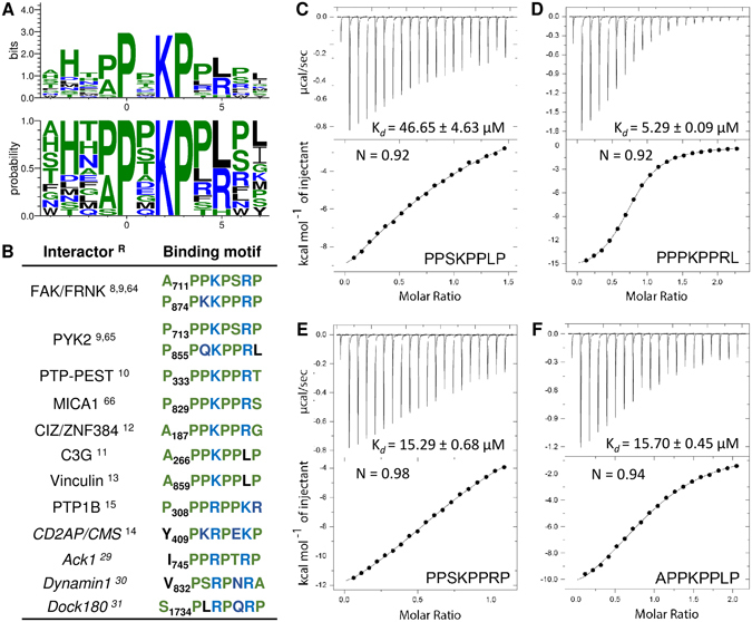
Figure 3.

(A) The CAS SH3 binding motif based on the 14 unique sequences obtained from phage display. The x-axis shows the residue position relative to proline (position 0)64–66. (B) The CAS SH3 domain binding interaction partners with their respective binding motifs. Interactors with small differences in binding motif are in italics. References (R) are superscripted. (C–E) Isothermal titration calorimetry (ITC) data obtained for the interaction of CAS SH3 with four synthetic peptides.
