Figure 1.

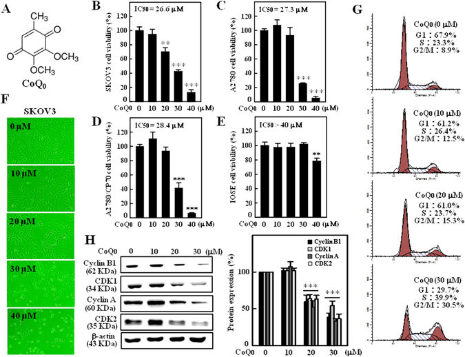
CoQ0 inhibits growth of human ovarian carcinoma cells and induces G2/M cell-cycle arrest in SKOV-3 cells. (A) Structure of CoQ0. (B–D) Human ovarian carcinoma (SKOV-3, A2870 and A2870/CP-70) and (E) human ovarian surface epithelial (IOSE) cells were treated with increasing concentrations of CoQ0 (0–40 µM) for 24 h. Cell viability was determined using MTT assay. (F) Morphological changes in CoQ0-treated (0–40 µM, 24 h) SKOV-3 cells were examined by phase-contrast microscope (200 × magnification). (G) SKOV-3 cells were treated with CoQ0 (0–30 µM) for 24 h, stained with PI and analyzed for cell-cycle phase using flow cytometry. The cellular distributions (percentage) in different phases of cell-cycle (G1, S and G2/M) were determined after CoQ0 treatment. Flow cytometry images shown here are from one representative analysis that was repeated three times with similar results. (H) SKOV-3 cells were treated with CoQ0 (0–30 µM) for 24 h, and cell-cycle regulatory proteins, cyclin B1, CDK1, cyclin A and CDK2 were examined using Western blot. Relative changes in protein intensities were quantified by commercially available software, and presented as histogram, control being as 1-fold. Results expressed as mean ± SD of three independent assays (n = 3), and significant at **p < 0.01; ***p < 0.001 compared with untreated control cells.
