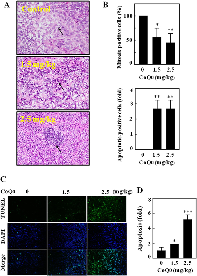
Figure 11.

Histopathological analyses of mitosis and apoptosis in the tumors of SKOV-3 xenografted nude mice followed by CoQ0 treatment. (A,B) Histopathological staining was performed to detect the mitotic- and apoptotic-cells in SKOV-3 xenografted tumors following CoQ0 (1.5 and 2.5 mg/kg) treatment using light microscopy (20 × and 200 × magnification). Arrows indicate mitotic (tumor control) and pyknotic tumor cells (CoQ0). The number of mitotic-positive and apoptotic-positive cells in microscopic fields of 3 samples was averaged, and presented as histogram. (C,D) In situ detection of apoptosis was performed using TUNEL staining in tumor sections from control and experimental analogues treated with CoQ0 (1.5 and 2.5 mg/kg). The number of TUNEL-positive cells were quantified from 3 microscopic fields, and averaged. Results expressed as mean ± SD of three independent assays (n = 3). Significant at *p < 0.05; **p < 0.01; ***p < 0.001 compared with vehicle treated mice.
