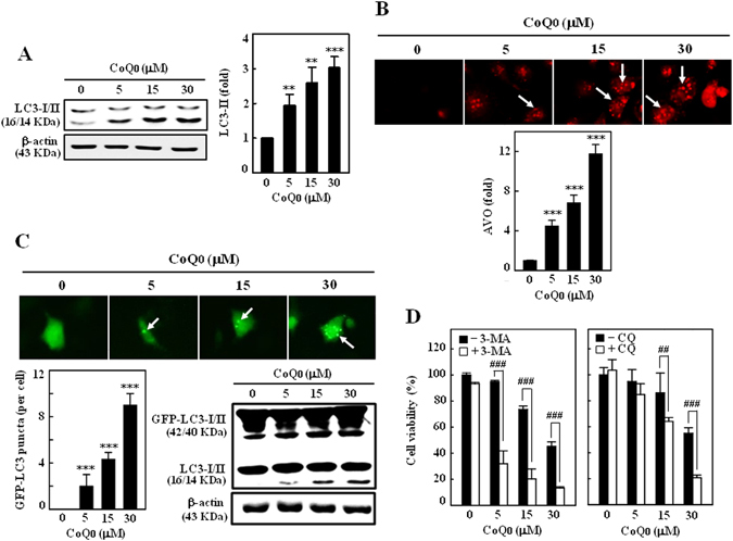
Figure 3.

CoQ0 promotes cytoprotective autophagy as a survival mechanism in SKOV-3 cells. (A) Cells were treated with various concentrations of CoQ0 (0–30 µM) for 24 h and then conversion of LC3-I to LC3-II was determined by Western blot. Relative changes in the intensities of protein bands were quantified by commercially available quantitative software. (B) CoQ0 induces AVOs formation. Cells were treated with CoQ0 (0–30 µM) for 24 h and stained with AO. Formation of AVOs, represented by red fluorescence intensity (in lysosomes) was visualized under a red filter fluorescence microscope (100 × magnification). Number of AO stained cells was presented as histogram, control being as 1.0 fold. (C) CoQ0 promotes conversion of GFP-LC3. Cells were transfected with GFP-LC3 expression vector for 24 h, and then treated with CoQ0 (0–30 µM) for 24 h. GFP-LC3 dots in cells were observed under a confocal microscope (200 × magnification). Conversions of GFP-LC3 and endogenous LC3 were determined by Western blot. (D) Cells were treated with autophagy inhibitors (2 mM 3-MA or 10 μM CQ) for 1 h followed by CoQ0 (0–30 µM) for 24 h, and viability was assayed by MTT assay. Results expressed as mean ± SD of three independent assays (n = 3). Significant at **p < 0.01; ***p < 0.001 compared with untreated control, and significant at ## p < 0.01; ### p < 0.001 compared with CoQ0 alone treated cells.
