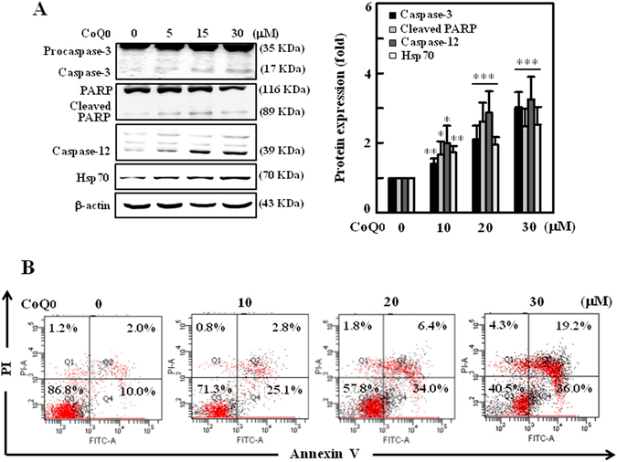
Figure 4.

CoQ0 induces apoptosis through mitochondrial and ER stress pathways in SKOV-3 cells. (A) Cells were treated with CoQ0 (0–30 µM) for 24 h, and apoptotic proteins involved in mitochondrial pathway (procaspse-3, caspase-3 and PARP) and ER stress pathway (caspase-12 and Hsp70) were estimated by Western blot. Changes in protein intensities were quantified by commercially available software, and presented as histogram. (B) Annexin-V-FITC and PI staining was used to identify the early/late apoptosis or necrosis of cells followed by CoQ0 (0–30 µM) treatment. Results from flow cytometry analysis in each quadrant (Q) are labeled and interpreted as follows: (Q1) PI positive, Annexin-V-FITC-negative stained cells/necrosis. (Q2) PI positive, Annexin-V-FITC-positive stained cells/late apoptosis. (Q3) cells negative for both PI and Annexin-V-FITC staining/normal live cells. (Q4) PI-negative, Annexin-V-FITC-positive stained cells/early apoptosis. Results expressed as mean ± SD of three independent assays (n = 3), and significant at *p < 0.05; **p < 0.01; ***p < 0.001 compared with untreated control cells.
