
Figure 2.
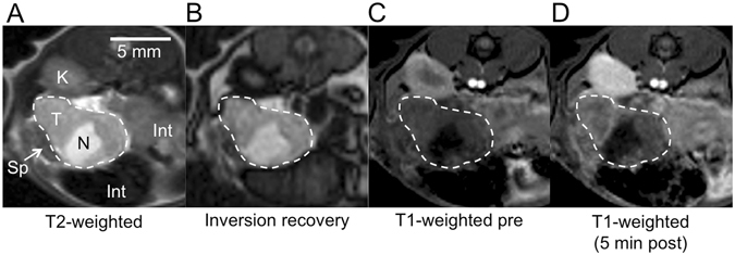
Axial MR images used to identify and characterize PDAC tumours in the mouse abdomen. Tumour is outlined. (A) T2-weighted image. T = tumour, N = necrosis, Sp = spleen, K = kidney, Int = intestine. (B) Pre-injection inversion recovery image (TR/TE/TI = 3200 ms/9.6 ms/487 ms). (C) Pre-injection T1-weighted image shows little contrast between tumour and surrounding tissue. (D) T1-weighted image at 5 min after injection of the collagen-targeted probe shows improved contrast and heterogeneous signal enhancement within the tumour.