Figure 3.

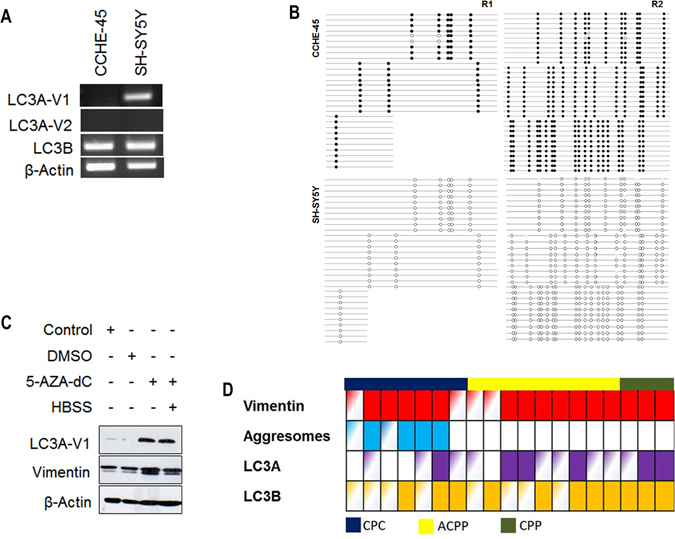
Inactivation of LC3A expression in choroid plexus carcinoma. (A) Expression of LC3A-V1, LC3A-V2 and LC3B were determined using RT-PCR in CCHE-45 and SH-SY5Y cell lines. β-actin was used as an internal control. (B) Bisulfite modified DNA PCR products from CCHE-45 and SH-SY5Y. Primers were designed to amplify CpG island (R1 and R2) upstream of LC3A-V1 using bisulfite sequencing. Diagrams show methylated CG dinucleotide in CCHE-45 R1 and R2 with no methylation detected in SH-SY5Y. (C) Western blot analysis of CCHE-45 cells treated with 10 µM 5-AZA-dC for 4 days then serum starved in HBSS for 2 hours. LC3A protein was restored following 5-AZA-dC treatment. No change in vimentin protein levels was detected. (D) Schematic representation for immunostaining of 19 cases CPC (blue), ACPP (yellow) or CPP (olive). FFPE tissue sections were stained with vimentin, LC3A or LC3B. Solid, partial and clear color indicates positive, focal or negative stain respectively.
