Figure 2.

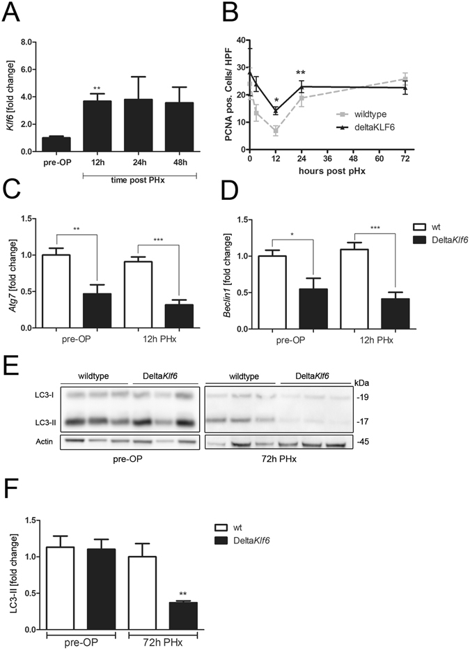
KLF6 affects liver regeneration and expression levels of autophagy-related genes after partial hepatectomy (PHx). Klf6 expression levels were determined by qRT-PCR in mouse liver tissue before (pre-OP) and 12 h, 24 h or 48 h after 70% partial hepatectomy (PHx, n = 6/group) (A). Cell proliferation was assessed by quantification of PCNA positive cells in liver tissue of wildtype (wt) and DeltaKlf6 mice 72 h after PHx (B). Expression-levels of Atg7 (C) and Beclin1 (D) were measured by qRT-PCR in liver tissue of wt and DeltaKlf6 mice before and 12 h after PHx. Autophagy was assessed by LC3 Western blotting and quantified by densitometry of specific protein bands (E,F) in liver tissue of wt and DeltaKlf6 mice 72 h after PHx. Shown are representative Western blot images (E) and densitometric quantification of LC3-II-bands normalized to loading control beta-Actin (F); fold change versus control shown as mean ± SEM of n = 6 mice per group; full length Western blot images are given in Supplementary information). *Represents p-value < 0.05 and **Indicates p-value < 0.01 as assessed by 2-way ANOVA comparing wt mice with DeltaKlf6 animals at the same time point after PHx.
